Functional Imaging – Application of molecular imaging to assess tumor response to antiangiogenic and Notch inhibition therapies
Molecular Imaging of Therapeutic Response
Juan Rojas – Application of molecular imaging to assess tumor response to antiangiogenic and Notch inhibition therapies
It is important to accurately track the response of cancer to different treatments in order to maximize efficacy. Change in tumor volume is the clinical gold standard for assessing treatment response. However, functional variations can occur much earlier than measurable volume changes. Contrast-Enhanced Ultrasound (CEUS) is an important tool for assessing tumor progression and response to therapy since it can monitor functional changes in the physiology. Targeting ligands can be added to the contrast agent shell so that the agents only bind to the desired biomarker in a technique called Ultrasound Molecular Imaging (USMI). The returned ultrasound signal corresponds only to areas where the biomarker is expressed so USMI can be used to assess response to therapy by tracking changes in vascular biomarker expression due to different therapeutics.
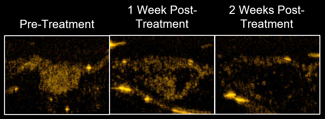
A recent study where mice bearing clear-cell Renal Cell Carcinoma were treated with anti-angiogenic therapy demonstrated that USMI can detect response to therapy earlier than changes in tumor volume in the treatment group compared to the control group. Furthermore, the results showed that response to the therapy was detected in over 90 % of cases one week after the start of treatment using USMI, while changes in tumor volume only detected response in 40 % of individual tumors. CD31 immunostaining was performed, and the results closely matched the USMI results, confirming that USMI can provide physiologically accurate information about disease state and response to therapy.
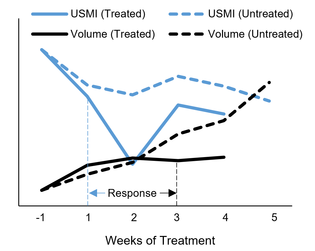
Fig 1- Individual response to treatment
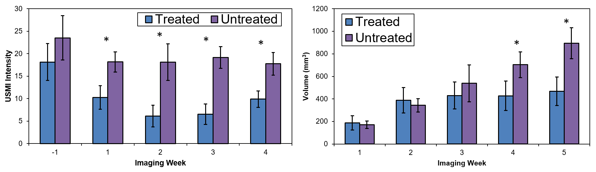
Fig 2- Group response to treatment