Functional Imaging – NC State collaboration Dog Imaging
Molecular Imaging of Therapeutic Response
Rachel White
Ultrasound molecular imaging is a method for non-invasively monitoring tumor response to therapeutic treatment, where image intensity corresponds to biomarker expression. Microbubbles can be targeted, via attachment of ligands and antibodies, to specific cellular structures and therefore function as molecular probes. Since cellular and molecular changes occur before noticeable changes in tumor volume, molecular imaging provides faster and more accurate feedback on therapeutic response. Furthermore, acoustic radiation force has been shown to increase the binding efficiency of targeted microbubbles, enhancing molecular imaging. Currently, we are using these techniques to monitor the response to therapy in canines with soft tissue sarcomas. Specifically, bubbles targeted to VEGFR2 and αvβ3 integrin, angiogenic markers overexpressed in the tumor microenvironment, are being used to track changes in the tumor microenvironment. This is in collaboration with the North Carolina State Veterinary School, the Borden group at the University of Colorado – Boulder, and the Colorado State University Veterinary Hospital.
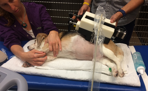
Figure 1: Testing the imaging system

Figure 2: Acoustic radiation force resulting in microbubble displacement in a 200 um tube.