Functional Imaging – Acoustic Angiography
Microvascular Imaging for Assessment of Tumor Angiogenesis
Sarah Shelton
Pre-Clinical Study
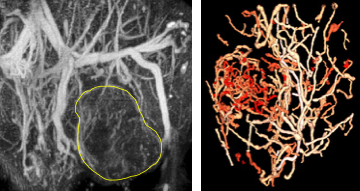
Acoustic angiography imaging relies on the production of superharmonics, a microbubble-specific phenomenon. By transmitting at a low frequency and receiving at a much higher frequency we isolate contrast signal with high resolution and clarity. These 3D angiography images can be acquired in only 1-2 minutes, resolving vessels as small as 100-150 μm in diameter to a depth of approximately 2 cm. Our research seeks to analyze vascular geometry (density, tortuosity, etc.) for tumor detection and diagnosis. Published work in pre-clinical models indicated that the tortuosity induced by a tumor extends beyond the margin of the tumor itself and that small 2 mm tumors can be distinguished based on their unique vascular patterns using simple methodology. We seek to address biological and clinical questions about microvascular remodeling using this unique imaging approach, and ongoing projects aim to improve the sensitivity and maximum imaging depth of this technique.
Clinical Study
We are also working towards clinical translation of acoustic angiography for the characterization of breast lesions. Many patients with suspicious breast lesions identified by mammography ultimately undergo a biopsy to confirm diagnosis, and the majority of these biopsies are negative for disease. Therefore, we aim to evaluate the value of acoustic angiography imaging for diagnosis of breast cancer, with potential for avoiding unnecessary biopsies in the future.

