
Functional Imaging – Application of contrast enhanced perfusion imaging to assess renal diseases
Perfusion Imaging
Sunny Kasoji – Application of contrast enhanced perfusion imaging to assess renal diseases
One application of contrast enhanced ultrasound imaging is perfusion imaging, or the dynamic imaging of blood flow. As a strictly intravascular blood-pooling agent, microbubble contrast agents are effective at representing blood flow. Tissue perfusion has been shown to be affected by both disease and therapy. For example, growing solid tumors require increased vasculature due to increased metabolic demand which can result in increased blood flow. Alternatively, a diseased tissue may undergo inflammation which results in increased perfusion. In research, perfusion imaging with CEUS has been used to as a method for diagnosing tumor malignancy, assessing response to therapy, or assessing organ function.
In the Dayton Lab, we use perfusion imaging in many different pre-clinical applications ranging from assessing changes in kidney function in a pre-diabetic rat kidney model to assessing tumor response to novel therapies. In all cases the goal is to improve the efficacy of diagnosis by providing early detection of disease and early assessment of therapy for improving patient outcomes.
Recently, we have initiated several clinical studies that are investigating the use of perfusion imaging for assessing kidney health.
- Renal Cell Carcinoma (RCC): RCC affects approximately 1 in every 100 American individuals, with 62,000 new cases estimated to be diagnosed in 2015. Traditional gray-scale ultrasound is effective in detecting solid renal lesions, but cannot further characterize them. Contrast CT and MRI are the standard imaging exams used to assess the malignancy of solid masses and to characterize perfusion patterns, however the contrast sensitivity does not adequately resolve histological characteristics of many types of tumors arising in the kidney, and the nephrotoxicity of the contrast agents often severely limits their use. Recent biomedical research with contrast-enhanced ultrasound (CEUS), a non-toxic and non-ionizing imaging modality, has shown evidence of the ability to characterize renal lesions based on qualitative enhancement and perfusion patterns; however few studies have explored quantitative approaches to assessing these characteristics. We quantified perfusion patterns in 16 patients diagnosed with renal masses and correlated specific metrics with pathological findings.
- Chronic kidney disease (CKD): CKD affects over 8 million people in the United States and is a major risk factor for cardiovascular disease, hospitalization and death. Diagnosis is usually made with blood and urine tests, and imaging is used primarily to rule out obstruction or determine size. Blood and urine tests are also used to follow disease progression with imaging used only for specific situations. In this ongoing clinical study, we evaluate the feasibility of using microbubble contrast agents to perform contrast-enhanced ultrasound (CEUS) and develop qualitative and quantitative measures of perfusion in patients with CKD. CEUS imaging may provide information about kidney perfusion that will aid in early diagnosis, disease progression and response to therapy.
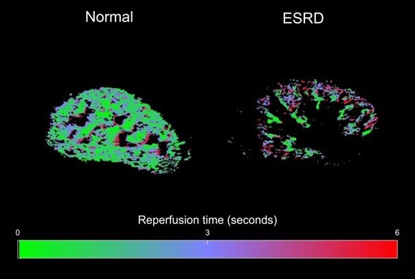
Figure 1. Reperfusion maps of renal parenchyma for healthy volunteer (left) and CKD patient (right). ESRD – end stage renal disease.
- Viscoelastic Response (VisR): In Progress