
Technology – PCCA
Phase Change Contrast Agents
Juan Rojas
Low boiling-point Phase Change Contrast Agents (PCCAs) have a liquid perfluorocarbon core and are smaller than conventional microbubble contrast agents which makes them able to circulate for longer in the vasculature and escape the vasculature in tumors and areas of inflammation. Due to their liquid core, PCCAs are not echogenic but can be vaporized into echogenic microbubbles using ultrasound pressures that are safe for diagnostic imaging. Therefore, PCCAs can generate on-demand contrast and can be used for imaging both inside and outside of the vasculature. Additionally, PCCAs are made by condensing microbubbles so modifications to the formulation, such as adding targeting ligands, can be easily be made on the precursor microbubbles.
PCCAs have been used extensively in vitro, but custom pulse sequences are required to properly use PCCAs in vivo for diagnostic imaging. The Verasonics Ultrasound Research system is a fully programmable system that allows for the development of new pulse sequences that can be tailored for PCCA use. Activation Pressure Matching (APM) is a technique developed for the Verasonics to deliver activation pulses that have the same pressure at every vaporization location regardless of depth by accounting for tissue attenuation and beam characteristics of the transducer such as elevational focusing due to a lens. APM produces uniform activation in a region of interest which is important for correct assessment of physiological characteristics in tissue such as biomarker expression and perfusion:
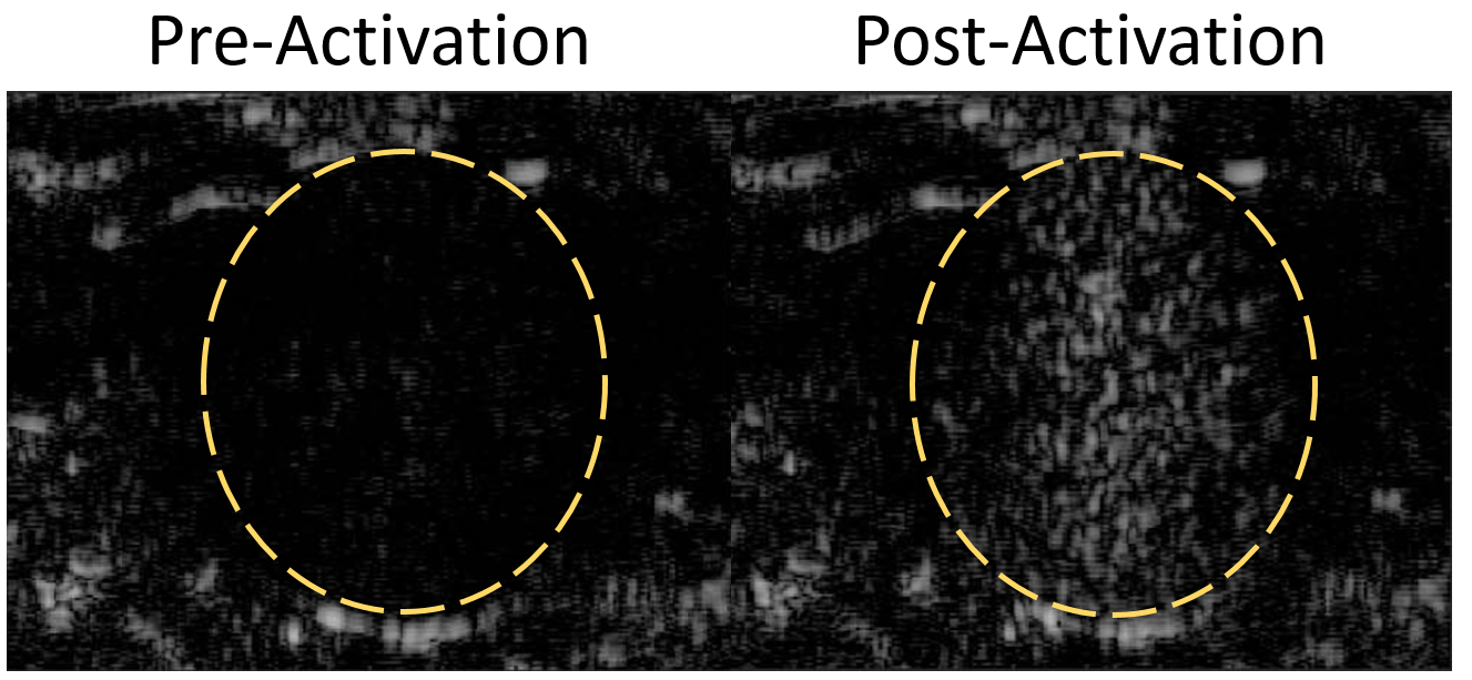 Fig 1- Rat kidney before and after vaporization of PCCAs
Furthermore, PCCA vaporization produces very unique acoustic signatures that are very different from microbubble or tissue echoes, and range in frequency between .25 and 2.5 MHz regardless of the activation pulse frequency. By using a transmit high/receive low strategy, droplet vaporization signals can be isolated to produce ultrasound images with high contrast-to-tissue (CTR) ratios. This technique has been demonstrated using 2 piston transducers, but It has recently been implemented using a single linear array transducer to image a rat kidney.
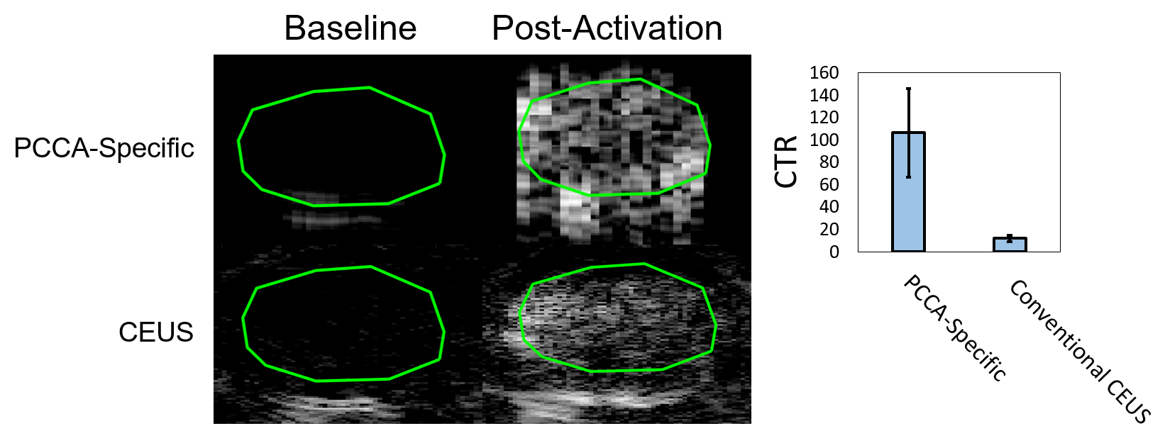
Fig 2- PCCA-specific imaging vs conventional contrast imaging.
The results showed that while the PCCA-specific images have a worse resolution, due to the low frequencies, they have a CTR that is significantly higher than images obtained by using conventional contrast images to capture the contrast generated by vaporization, as has been commonly done.